• Blog
  • About
  • Contact
  • Blog
  • About
  • Contact
Arianna Dolbee

All It Is Best To Know About Heel Discomfort

7/2/2017

 
Overview

Painful Heel

Heel pain is one of the most common conditions treated by podiatrists. It is often a message that something is in need of medical attention. Pain that occurs right after an injury or early in an illness may play a protective role, often warning us about the damage we have suffered. Heel pain is a problem which affects people of all ages and vocations, whether they are active or not and it comes in many different forms. Heel pain can also occur in children usually between the ages of 8 and 13, as they become increasingly active in sporting activities and during the growing phase.

Causes

Age plays a large role in the development of heel pain, particularly among those over 40. Being active is also a common factor of heel pain. Over time, the elasticity of the tissue in our feet decreases with age, causing us to become prone to damage and also slowing the body's ability to heal damage. Adolescents are also not immune to heel pain. Those who are active in sports are particularly prone to excessively stretching or straining the plantar fascia or Achilles tendon, causing severe heel pain. In most cases, heel pain develops in only one heel. There are many risk factors that lead to heel pain. Abnormal gait and excessive, repetitive stress are common factors in the development of pain and damage. Among the other risk factors involved with the development of heel pain are repetitive exercise or activities, such as long distance running or jumping from activities such as basketball. Obesity. Walking barefoot on hard surfaces. Prolonged standing. Wearing poor fitting shoes, or shoes that do not provide enough support or cushioning. Not stretching properly or at all before and after exercise. Those who are on their feet for long periods of time.

Symptoms

Pain in the bottom of the heel is the most common symptom. The pain is often described as a knife-like, pinpoint pain that is worse in the morning and generally improves throughout the day. By the end of the day the pain may be replaced by a dull ache that improves with rest. The pain results from stretching the damaged tissues. For the same reason atheletes' pain occurs during beginning stages of exercise and is relieved over time as warm-up loosens the fascia. Plantar fasciitis onset is usually gradual, only flaring up during exercise. If pain is ignored, it can eventually interfere with walking and overall, plantar fasciitis accounts for about ten percent of all running injuries.

Diagnosis

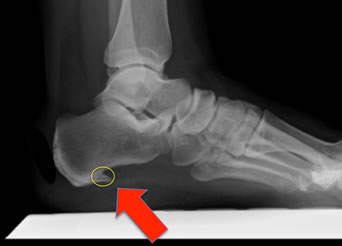
To arrive at a diagnosis, the foot and ankle surgeon will obtain your medical history and examine your foot. Throughout this process the surgeon rules out all the possible causes for your heel pain other than plantar fasciitis. In addition, diagnostic imaging studies such as x-rays or other imaging modalities may be used to distinguish the different types of heel pain. Sometimes heel spurs are found in patients with plantar fasciitis, but these are rarely a source of pain. When they are present, the condition may be diagnosed as plantar fasciitis/heel spur syndrome.

Non Surgical Treatment

Once diagnosed, treatment for plantar fasciitis may include one or more of the following: advice on footwear, in particular use of arch-supportive footwear; avoid walking barefoot; stretching exercises, shoe modifications such as heel pads, taping and strapping, anti-inflammatories and orthotic devices to correct abnormal foot mechanics. Injection therapy with corticosteroids is only advisable if all the conservative treatment methods mentioned above have been exhausted due to undesired effects implicated with steroid infusion in the heels.

Surgical Treatment

Although most patients with plantar fasciitis respond to non-surgical treatment, a small percentage of patients may require surgery. If, after several months of non-surgical treatment, you continue to have heel pain, surgery will be considered. Your foot and ankle surgeon will discuss the surgical options with you and determine which approach would be most beneficial for you. No matter what kind of treatment you undergo for plantar fasciitis, the underlying causes that led to this condition may remain. Therefore, you will need to continue with preventive measures. Wearing supportive shoes, stretching, and using custom orthotic devices are the mainstay of long-term treatment for plantar fasciitis.

Why do I have pain in my heel?

Prevention

Pain In The Heel

Make sure you wear appropriate supportive shoes. Don't over-train in sports. Make sure you warm up, cool down and undertake an exercise regime that helps maintain flexibility. Manage your weight, obesity is a factor in causing plantar fasciitis. Avoid walking and running on hard surfaces if you are prone to pain. You should follow the recognized management protocol "RICED" rest, ice, compression, elevation and diagnosis. Rest, keep off the injured ankle as much as possible. Ice, applied for 20 minutes at a time every hour as long as swelling persists. Compression, support the ankle and foot with a firmly (not tightly) wrapped elastic bandage. Elevation, keep foot above heart level to minimize bruising and swelling. Diagnosis. Consult a medical professional (such as a Podiatrist or doctor) especially if you are worried about the injury, or if the pain or swelling gets worse. If the pain or swelling has not gone down significantly within 48 hours, also seek treatment. An accurate diagnosis is essential for proper rehabilitation of moderate to severe injuries.

A Way To Diagnose Fallen Arches

7/2/2017

 
Overview

Flat Foot

A ?fallen arch? is a change in the shape of the foot. The normal gap between the floor and the arch on the inside of the foot narrows. It is usually painful, sometimes cripplingly painful and crippling. The heel leans to the inside, giving the inner ankle a bowed appearance. The joints of the outside of the crush together causing pain and swelling. The ligaments on the inside of the ankle stretch causing additional pain. Over time, the joints of the midfoot and forefoot adapt to the tilting of the ankle so that the front of the foot remains on the floor, complicating the deformity.

Causes

There are many reasons why flat feet develop. Here?s a look at some of the most common causes. Genetics, weak arches, injury, arthritis, diabetes, age, wear and tear on feet, tibialis posterior (ruptured tendon). Nervous system or muscle diseases such as cerebral palsy. Weakness and tightness of other muscles and tendons higher up in the lower extremity. The way our arches form depends on several factors. Our feet are complex structures that comprise twenty-six bones, thirty-three joints, and more than 100 muscles, tendons, and ligaments each. Each foot forms two arches. The arch that runs from the heel to the toe is known as the longitudinal arch, while the one that runs the width is known as the transverse arch. Ligaments (fibrous tissues) give our arches their shape and hold our bones together. The plantar fascia (the long, strong band of connective tissue that runs along the sole of your foot) and muscles add secondary support. There are also foot pads that absorb impact and assist with weight-bearing functions. How these things intertwine and work together determines the formation of our arches. A structural abnormality or injury to one of these components can result in flatfoot.

Symptoms

Feet tire easily and become painful and achy, especially around the arch, ankle and heel. Swelling on the inside bottom of your feet. Back and leg pain. Difficulty standing on toes.

Diagnosis

Your doctor examines your feet to determine two things, whether you have flat feet and the cause or causes. An exam may include the following steps, Checking your health history for evidence of illnesses or injuries that could be linked to flat feet or fallen arches, Looking at the soles of your shoes for unusual wear patterns, Observing the feet and legs as you stand and do simple movements, such as raising up on your toes, Testing the strength of muscles and tendons, including other tendons in the feet and legs, such as the Achilles tendon or the posterior tibial tendon, Taking X-rays or an MRI of your feet.

high arch feet

Non Surgical Treatment

Most patients can be treated without surgery using orthotics, supportive shoes and braces. Because of the progressive nature of PTTD, early treatment is advised. If treated early enough, your symptoms may resolve without the need for surgery and progression of your condition can be arrested. In contrast, untreated PTTD could leave you with an extremely flat foot, painful arthritis in the foot and ankle, and increasing limitations on walking, running, or other activities.

Surgical Treatment

Flat Foot

This is rare and usually only offered if patients have significant abnormalities in their bones or muscles. Treatments include joint fusion, reshaping the bones in the foot, and occasionally moving around tendons in the foot to help balance out the stresses (called tendon transfer). Flat feet and fallen arches are common conditions that are in most cases asymptomatic. However, in patients who do have symptoms, treatments are available that can help reduce pain and promote efficient movement. Orthotic devices are well recognised as an excellent treatment and podiatrists can offer these different treatment modalities as individualised treatments for patients.

Prevention

Flatfeet in children are often an inherited family trait, but it may be possible to prevent the condition in some cases. Recent research has shown that there are several social or cultural factors that can cause flatfeet. These factors include the following, obesity, overweight, unnecessary orthopedic treatments, wearing rigid shoes at a young age, In 1992, a study in India of 2300 children aged 4-13 demonstrated a significant difference in the rate of flatfeet among those who wore shoes regularly and those who did not. In this study, wearing inflexible, closed-toe shoes in early childhood was shown to have a negative effect on the normal development of arches. Children who were allowed to go barefoot or who wore light sandals and slippers had a much lower rate of flatfeet. In 1999, a study in Spain of 1181 children aged 4-13 revealed that the use of orthopedic shoes for treatment of flatfeet in children not only failed to correct the problem, but actually worsened the condition by preventing the normal flexing and arch development of bare or lightly protected feet. Finally, in 2006, a study of 835 children aged 3-6 showed significant differences in the rate of flatfeet based on weight, with normal-weight children having lower rates of flatfeet than children who were overweight or obese. Among adults, flatfeet due to injury, disease, or normal aging are not preventable. However, when flatfeet are related to lifestyle factors, such as physical activities, shoe selection, and weight gain, careful attention to these factors may prevent the development of flatfeet.

Functional Leg Length Discrepancy Running

6/27/2017

 
Overview

Large leg length inequalities can be treated by staged lengthenings or by simultaneous ipsilateral femoral and tibial lengthenings. Additionally, lengthenings can be combined with appropriately timed epiphysiodesis in an effort to produce leg length equality. Staged lengthenings are often used for congenital deficiencies such as fibular hemimelia, in which 15 cm or more may be needed to produce leg length equality. We typically plan for the final lengthening to be completed by age 13 or 14 years, and allow at least 3 years between lengthenings. Lengthening of both the tibia and femur simultaneously requires aggressive therapy and treatment of soft tissue contractures. Curran et al[57] reported the need for surgical release of soft tissue contractures in 3 of 8 patients treated with simultaneous ipsilateral femoral and tibial lengthenings. Lengthening over an IM nail can be done in an effort to decrease the amount of time the fixator needs to be worn and to prevent angular malalignment. This technique requires that the patient be skeletally mature and it carries a higher risk of osteomyelitis (up to 15%). Additionally, if premature consolidation occurs, a repeat corticotomy is more difficult.Leg Length Discrepancy

Causes

Some limb-length differences are caused by actual anatomic differences from one side to the other (referred to as structural causes). The femur is longer (or shorter) or the cartilage between the femur and tibia is thicker (or thinner) on one side. There could be actual deformities in one femur or hip joint contributing to leg length differences from side to side. Even a small structural difference can amount to significant changes in the anatomy of the limb. A past history of leg fracture, developmental hip dysplasia, slipped capital femoral epiphysis (SCFE), short neck of the femur, or coxa vara can also lead to placement of the femoral head in the hip socket that is offset. The end-result can be a limb-length difference and early degenerative arthritis of the hip.

Symptoms

Back pain along with pain in the foot, knee, leg and hip on one side of the body are the main complaints. There may also be limping or head bop down on the short side or uneven arm swinging. The knee bend, hip or shoulder may be down on one side, and there may be uneven wear to the soles of shoes (usually more on the longer side).

Diagnosis

The doctor carefully examines the child. He or she checks to be sure the legs are actually different lengths. This is because problems with the hip (such as a loose joint) or back (scoliosis) can make the child appear to have one shorter leg, even though the legs are the same length. An X-ray of the child?s legs is taken. During the X-ray, a long ruler is put in the image so an accurate measurement of each leg bone can be taken. If an underlying cause of the discrepancy is suspected, tests are done to rule it out.

Non Surgical Treatment

Structural leg length discrepancy can be treated with a heel lift in the shorter leg?s shoe, if the leg length is greater than 5 mm. The use and size of the heel lift is determined by a physical therapist based on how much lift is needed to restore proper lumbopelvic biomechanics. In certain cases, surgical intervention may be needed to either shorten or lengthen the limb. An important component to any surgical procedure to correct leg length discrepancies is physical therapy. Physical therapy helps to stretch muscles and maintain joint flexibility, which is essential in the healing process. For a functional leg length discrepancy no heel lift is required, but proper manual therapy techniques and specific therapeutic exercise is needed to treat and normalize pelvic and lower extremity compensations. The number of treatments needed to hold the pelvis in a symmetrical position is different for each patient based on their presentation and biomechanical dysfunctions in their lower back, pelvis, hip, knee, and foot/ankle. If you have pain in your lower back or lower extremity and possibly a length discrepancy; the two symptoms could be related. A good place to start would be a physical therapy evaluation to determine whether you have a leg length discrepancy and if it could be contributing to your lower back pain, hip pain, knee pain, or leg pain.

LLD Insoles

how to grow taller at 16

Surgical Treatment

Surgery is another option. In some cases the longer extremity can be shortened, but a major shortening may weaken the muscles of the extremity. In growing children, lower extremities can also be equalized by a surgical procedure that stops the growth at one or two sites of the longer extremity, while leaving the remaining growth undisturbed. Your physician can tell you how much equalization can be attained by surgically halting one or more growth centers. The procedure is performed under X-ray control through very small incisions in the knee area. This procedure will not cause an immediate correction in length. Instead, the LLD will gradually decrease as the opposite extremity continues to grow and "catch up." Timing of the procedure is critical; the goal is to attain equal length of the extremities at skeletal maturity, usually in the mid- to late teens. Disadvantages of this option include the possibility of slight over-correction or under-correction of the LLD and the patient?s adult height will be less than if the shorter extremity had been lengthened. Correction of significant LLDs by this method may make a patient?s body look slightly disproportionate because of the shorter legs.

Addressing Mortons Neuroma

6/2/2017

 
Overview

Morton neuromaMorton's neuroma is the common name given to the nerve irritation that is found in the ball of the foot that may or may not be accompanied by an inter-metatarsal bursae (a bursa-neuromal complex). It is often associated with inflammation or degeneration and often occurs with constant pressure or irritation of the nerve from the surrounding bony structures or local bursas (fluid filled sacs). Morton's Neuroma can cause symptoms such as a sharp pain, burning even a lack of feeling in the ball of the foot and associated toes.

Causes

Some say that this condition should not be called Morton's neuroma as, in fact, it is not actually a neuroma. A neuroma is a non-cancerous (benign) tumour that grows from the fibrous coverings of a nerve. There is no tumour formation in Morton's neuroma. The anatomy of the bones of the foot is also thought to contribute to the development of Morton's neuroma. For example, the space between the long bones (metatarsals) in the foot is narrower between the second and third, and between the third and fourth metatarsals. This means that the nerves that run between these metatarsals are more likely to be compressed and irritated. Wearing narrow shoes can make this compression worse.

Symptoms

Patients will complain of numbness, a ?pins and needles? type of tingling and loss of sensation in the toes. Burning pain in the ball of the foot that may radiate into the toes. The pain generally intensifies with activity or wearing shoes. Night pain is rare. There may also be numbness in the toes, or an unpleasant feeling in the toes. Runners may feel pain as they push off from the starting block. High-heeled shoes, which put the foot in a similar position to the push-off, can also aggravate the condition. Tight, narrow shoes also aggravate this condition by compressing the toe bones and pinching the nerve.

Diagnosis

The most common condition misdiagnosed as Morton's neuroma is metatarsophalangeal (MTP) joint synovitis. When pain occurs in the third interspace, the clinician may misdiagnose the condition as Morton's neuroma instead of MTP synovitis, which may manifest very much like Morton's neuroma. MTP synovitis is distinguished from Morton's neuroma by subtle swelling around the joint, pain localized mainly within the joint, and pain with forced toe flexion. Palpation of the MTP joint is performed best with a pinching maneuver from the dorsal and plantar aspects of the joint to elicit tenderness of the joint. Other conditions often misdiagnosed as Morton's neuroma include the following. Stress fracture of the neck of the metatarsal. Rheumatoid arthritis and other systemic arthritic conditions. Hammertoe. Metatarsalgia (ie, plantar tenderness over the metatarsal head) Less common conditions that have overlapping symptoms with Morton's neuroma include the following. Neoplasms. Metatarsal head osteonecrosis. Freiburg. steochondrosis. Ganglion cysts. Intermetatarsal bursal fluid collections. True neuromas.

Non Surgical Treatment

Conservative treatment for Morton?s neuroma involves footwear that allows your forefoot to spread. High-heeled shoes cause neuromas by squeezing and stretching your involved intermetatarsal nerve across the ball of your foot and should be avoided as often as possible. A shoe that possesses any toe spring will also place more stress on your foot nerves and increase your likelihood of developing a neuroma. Test shoes before you buy them to see if they are appropriate for your feet. Select shoes that have a removable liner or insole, and stand on the liner, noting the position of your foot. If your foot is wider than your liner, that shoe will irritate your neuroma by squeezing your metatarsal bones together.Morton neuroma

Surgical Treatment

The above measures are often sufficient to resolve Morton?s Neuroma. Should the condition persist or worsen despite these efforts, surgery may be recommended to remove the Neuroma. The surgery requires only a short recovery period, though permanent numbness in the affected toes can result, so such surgery is generally used as a last resort.

Hammer Toe Cure

7/2/2015

 
Hammer ToeOverview
Hammer toes (hammertoe) is a deformity of the second, third, or fourth toes. In this condition, the toe is bent at the middle joint, into an upward position, causing it to resemble a hammer (sometimes decribed as ?curled toes?). Left untreated, hammer toes can become inflexible and require surgery. Toes which take on a curled appearance are hammer toes. Mallet toe is a similar condition, but affects the upper joint of a toe.

Causes
A common cause of hammer toe is wearing shoes that do not fit properly. Poorly-fitting shoes can hold the toes in an abnormal position and result in tightening of the muscles required to maintain that position. In particular, shoes that have high heels and are narrow at front tend to push the toes into an abnormal, bent position. Less commonly, diseases of the nerves, muscles, or joints (such as arthritis) can result in the hammer toe deformity. Hammer Toe

Symptoms
If the toes remain in the hammertoe position for long periods, the tendons on the top of the foot will tighten over time because they are not stretched to their full length. Eventually, the tendons shorten enough that the toe stays bent, even when shoes are not being worn. The symptoms of hammertoe include a curling toe, pain or discomfort in the toes and ball of the foot or the front of the leg, especially when toes are stretched downward, thickening of the skin above or below the affected toe with the formation of corns or calluses, difficulty finding shoes that fit well. In its early stages, hammertoe is not obvious. Frequently, hammertoe does not cause any symptoms except for the claw-like toe shape.

Diagnosis
The treatment options vary with the type and severity of each hammer toe, although identifying the deformity early in its development is important to avoid surgery. Your podiatric physician will examine and X-ray the affected area and recommend a treatment plan specific to your condition.

Non Surgical Treatment
Mild hammer toe in children can be treated by manipulating and splinting the affected toe. The following changes in footwear may help relieve symptoms. Wear the right size shoes or shoes with wide toe boxes for comfort, and to avoid making hammer toe worse. Avoid high heels as much as possible. Wear soft insoles to relieve pressure on the toe. Protect the joint that is sticking out with corn pads or felt pads. A foot doctor can make foot devices called hammer toe regulators or straighteners for you, or you can buy them at the store. Exercises may be helpful. You can try gentle stretching exercises if the toe is not already in a fixed position. PIcking up a towel with your toes can help stretch and straighten the small muscles in the foot.

Surgical Treatment
If you are unable to flex your toe, surgery is the only option to restore movement. Surgery is used to reposition the toe, remove deformed or injured bone, and realign your tendons. Surgery is normally done on an outpatient basis, so you can return home on the day of your surgery.

Working With Bunions

6/5/2015

 
Overview
Bunion Pain 
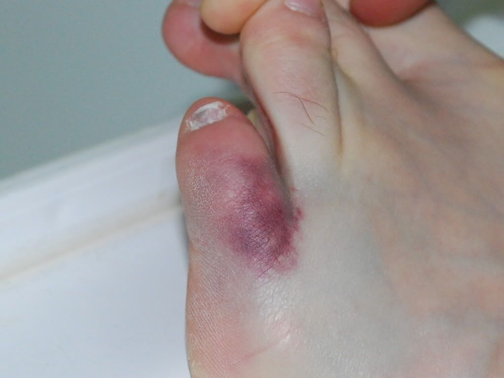
A bunion (hallux abducto valgus) is a bony lump or enlargement that forms in the joint (metatarsal phalangeal joint) at the base of the big toe. The big toe points toward the other toes (lateral deviation) as the bunion progresses. The joint and surrounding tissue becomes inflamed and painful. Occasionally bunions can also form at the joint at the base of the smallest (fifth) toe. They are called a tailor's bunion or bunionette. Bunions can be accompanied by bursitis (inflammation of a small fluid-filled sac adjacent to the joint). With an advanced bunion the big toe may be so deformed that it has to lie over or under the second toe. In this situation it hurts to walk or wear any kind of shoe. About one in three people in most Western countries will get a bunion. They are about ten times more common in women than in men. Older people are more often affected than younger people, although bunions do occur in children with misaligned feet. Bunions should not be confused with gout or arthritis, which can also cause inflammation, deformity and pain around the toes and feet.

Causes
Women traditionally have a higher rate of bunions, which is to be expected, since it is they who have traditionally worn shoes with high heels, a narrow toe box, or whatever fashion dictates from year to year. However, men can suffer from bunions as well, as can anyone for whom the right (or wrong) conditions exist, poor foot mechanics, improper footwear, occupational hazards, health and genetic predisposition. Finally, bunions have long been a condition associated with the elderly, and although they often appear in conjunction with inflammatory joint diseases such as arthritis (which is often associated with age), they can strike at any point in life, including adolescence.

Symptoms
Just because you have a bunion does not mean you have to have pain. There are some people with very severe bunions and no pain and people with mild bunions and a lot of pain. Symptoms for a bunion may include pain on the inside of your foot at the big toe joint. Swelling on the inside of your foot at the big toe joint. Redness on the inside of your foot at the big toe joint. Numbness or burning in the big toe (hallux). Decreased motion at the big toe joint. Painful bursa (fluid-filled sac) on the inside of your foot at the big toe joint. Pain while wearing shoes, especially shoes too narrow or with high heels. Joint pain during activities. Other conditions which may appear with bunions include corns in between the big toe and second toe. Callous formation on the side or bottom of the big toe or big toe joint. Callous under the second toe joint. Pain in the second toe joint.

Diagnosis
Generally, observation is adequate to diagnose a bunion, as the bump is obvious on the side of the foot or base of the big toe. However, your physician may order X-rays that will show the extent of the deformity of the foot.

Non Surgical Treatment
Many people with bunions are quite comfortable if they wear wide, well fitting shoes and give them time to adapt to the shape of their feet. A small pad over the bony prominence, which can be bought from a chemist or chiropodist, can take the pressure of the shoe off the bunion. High heels tend to squeeze the foot into the front of the shoe and should be avoided. It is often worthwhile seeing a chiropodist if these simple measures are not quite enough. Bunion Pain

Surgical Treatment
If non-surgical treatments fail to relieve bunion pain and when the pain of a bunion interferes with daily activities, it?s time to discuss surgical options with a foot and ankle surgeon. Together you can decide if surgery is best for you. A variety of surgical procedures is available to treat bunions. The procedures are designed to remove the ?bump? of bone, correct the changes in the bony structure of the foot, and correct soft tissue changes that may also have occurred. The goal of surgery is the reduction of pain. In selecting the procedure or combination of procedures for your particular case, the foot and ankle surgeon will take into consideration the extent of your deformity based on the x-ray findings, your age, your activity level, and other factors. The length of the recovery period will vary, depending on the procedure or procedures performed.

Learn About Over-Pronation Of The Foot

5/31/2015

 
Overview

Overpronation and underpronation describe general foot movements. These terms do not necessarily describe a medical problem with a foot. For example, you can overpronate and not have any problems or symptoms at all. It is important to have your foot structure and symptoms adequately assessed by your prescribing physician and a qualified practitioner such as a Canadian Certified Pedorthist. Once the underlying conditions and mechanical faults are assessed, an appropriate treatment plan including possible orthotic and footwear recommendations can be made.Overpronation

Causes

There may be several possible causes of over pronation. The condition may begin as early as birth. However, there are several more common explanations for the condition. First, wear and tear on the muscles throughout the foot, either from aging or repetitive strain, causes the muscles to weaken, thereby causing the foot to turn excessively inward. Also, standing or walking on high heels for an extended period of time also places strain and pressure on the foot which can weaken the tissue. Lastly, shoes play a very common factor in the development of over pronation. Shoes that fail to provide adequate support through the arch commonly lead to over pronation.

Symptoms

When standing, your heels lean inward. When standing, one or both of your knee caps turn inward. Conditions such as a flat feet or bunions may occur. You develop knee pain when you are active or involved in athletics. The knee pain slowly goes away when you rest. You abnormally wear out the soles and heels of your shoes very quickly.

Diagnosis

One of the easiest ways to determine if you overpronate is to look at the bottom of your shoes. Overpronation causes disproportionate wear on the inner side of the shoe. Another way to tell if you might overpronate is to have someone look at the back of your legs and feet, while you are standing. The Achilles tendon runs from the calf muscle to the heel bone, and is visible at the back of the ankle. Normally it runs in a straight line down to the heel. An indication of overpronation is if the tendon is angled to the outside of the foot, and the bone on the inner ankle appears to be more prominent than the outer anklebone. There might also be a bulge visible on the inside of the foot when standing normally. A third home diagnostic test is called the ?wet test?. Wet your foot and stand on a surface that will show an imprint, such as construction paper, or a sidewalk. You overpronate if the imprint shows a complete impression of your foot (as opposed to there being a space where your arch did not touch the ground).Pronation

Non Surgical Treatment

Flat feet and fallen arches can be treated effectively by wearing an orthotic insert in your shoes. Orthotics can be custom-made and prescribed by your foot specialist (podiatrist), or you can use a so called pre-made foot orthotic. Most people do not require expensive custom-made orthotics to combat excess pronation, unless they have a specific medical foot condition. Footlogics orthotic insoles were developed to correct excess pronation, thereby providing sustainable, long-lasting pain relief to many aches and pains in a natural way. Footlogics Comfort, Casual and Sports are products which promote excellent biomechanical control of the foot.

Surgical Treatment

Subtalar Arthroereisis. Primary benefit is that yje surgery is minimally invasive and fully reversible. the primary risk is a high chance of device displacement, generally not tolerated in adults.

An implant is pushed into the foot to block the excessive motion of the ankle bone. Generally only used in pediatric patients and in combination with other procedures, such as tendon lengthening. Reported removal rates vary from 38% - 100%, depending on manufacturer.

What Exactly Is Severs Disease?

5/17/2015

 
Overview

Heel pain is common in children. While it can occur after a specific injury, it is also commonly caused by Sever's disease, a type of overuse syndrome, like shin splints or Osgood-Schlatter?s disease. Children with Sever's disease, which is also called calcaneal apophysitis, develop inflammation where the Achilles tendon inserts at the calcaneus, or heel bone. This inflammation causes pain, which can vary depending on the type of activity your child is doing, and is generally worse after activity(such as running and jumping) and improves with rest. Sometimes squeezing the heel can cause pain and occasionally it can be felt under the heel.

Causes

This condition most commonly occurs due to repetitive or prolonged activities placing strain on the heel's growth plate, typically during a period of rapid growth. These activities (or sports) usually involve excessive walking, running, jumping or hopping. Severs disease may also be more likely to occur following a poorly rehabilitated sprained ankle, in patients with poor foot biomechanics or those who use inappropriate footwear. In young athletes, this condition is commonly seen in running and jumping sports, such as football, basketball, netball and athletics.

Symptoms

Chief complaint is heel pain which increases pain during running and jumping activities. Pain is localized to the very posterior aspect of the heel. Pain is elicited only with weightbearing. Mild involvement is present if pain is brought on only with running during sports. The symptoms can be severe, with pain (and possibly limp) with activities of daily living (ie walking).

Diagnosis

To diagnose the cause of the child?s heel pain and rule out other more serious conditions, the foot and ankle surgeon obtains a thorough medical history and asks questions about recent activities. The surgeon will also examine the child?s foot and leg. X-rays are often used to evaluate the condition. Other advanced imaging studies and laboratory tests may also be ordered.

Non Surgical Treatment

For patients suffering from Sever's disease, the pediatric orthopaedic surgeon will often recommend a conservative treatment plan including anti-inflammatory medication (as directed by the doctor). Application of ice to the heel. Calf, hamstring, and heel stretches. Orthotics. Modification of activities. There are rarely any complications with the treatment of Sever's disease, and symptoms generally resolve within 2 weeks to 2 months. Patients can typically return to playing sports again after the heel pain has resolved. The physician will let confirm when it is safe to resume physical activities.

Exercise

The following exercises are commonly prescribed to patients with Severs disease. You should discuss the suitability of these exercises with your physiotherapist prior to beginning them. Generally, they should be performed 1 - 3 times daily and only provided they do not cause or increase symptoms. Your physiotherapist can advise when it is appropriate to begin the initial exercises and eventually progress to the intermediate, advanced and other exercises. As a general rule, addition of exercises or progression to more advanced exercises should take place provided there is no increase in symptoms. Calf Stretch with Towel. Begin this stretch in long sitting with your leg to be stretched in front of you. Your knee and back should be straight and a towel or rigid band placed around your foot as demonstrated. Using your foot, ankle and the towel, bring your toes towards your head as far as you can go without pain and provided you feel no more than a mild to moderate stretch in the back of your calf, Achilles tendon or leg. Hold for 5 seconds and repeat 10 times at a mild to moderate stretch provided the exercise is pain free. Calf Stretch with Towel. Begin this exercise with a resistance band around your foot and your foot and ankle held up towards your head. Slowly move your foot and ankle down against the resistance band as far as possible and comfortable without pain, tightening your calf muscle. Very slowly return back to the starting position. Repeat 10 - 20 times provided the exercise is pain free. Once you can perform 20 repetitions consistently without pain, the exercise can be progressed by gradually increasing the resistance of the band provided there is no increase in symptoms. Bridging. Begin this exercise lying on your back in the position demonstrated. Slowly lift your bottom pushing through your feet, until your knees, hips and shoulders are in a straight line. Tighten your bottom muscles (gluteals) as you do this. Hold for 2 seconds then slowly lower your bottom back down. Repeat 10 times provided the exercise is pain free.

Achilles Rupture Fallacies

4/30/2015

 
Overview
Achilles Tendinitis Complete Achilles tendon ruptures occur most commonly at the mid-substance, but also distally at the insertion site or proximally at the myotendinous junction. These can be traumatic and devastating injuries, resulting in significant pain, disability, and healthcare cost. As many as 2.5 million individuals sustain Achilles tendon ruptures each year and the incidence is rising. This trend is due, in part, to an increase in athletic participation across individuals of all ages.

Causes
A rupture occurs when a patient overstretches the Achilles tendon, an act which causes it to tear partially or completely. Achilles tendon ruptures can occur during athletic play or any time the tendon is stretched in an unexpected way.

Symptoms
Although it's possible to have no signs or symptoms with an Achilles tendon rupture, most people experience pain, possibly severe, and swelling near your heel, an inability to bend your foot downward or "push off" the injured leg when you walk, an inability to stand on your toes on the injured leg, a popping or snapping sound when the injury occurs. Seek medical advice immediately if you feel a pop or snap in your heel, especially if you can't walk properly afterward.

Diagnosis
During the physical exam, your doctor will inspect your lower leg for tenderness and swelling. In many cases, doctors can feel a gap in your tendon if a complete rupture has occurred. Achilles tendon rupture can be diagnosed reliably with clinical examination, but if there?s a question about the extent of your Achilles tendon injury then your doctor may order a magnetic resonance imaging (MRI) scan.

Non Surgical Treatment
The best treatment for a ruptured Achilles tendon often depends on your age, activity level and the severity of your injury. In general, younger and more active people often choose surgery to repair a completely ruptured Achilles tendon while older people are more likely to opt for nonsurgical treatment. Recent studies, however, have shown fairly equal effectiveness of both operative and nonoperative management. Nonsurgical treatment. This approach typically involves wearing a cast or walking boot with wedges to elevate your heel; this allows the ends of your torn tendon to heal. This method can be effective, and it avoids the risks, such as infection, associated with surgery. However, the likelihood of re-rupture may be higher with a nonsurgical approach, and recovery can take longer. If re-rupture occurs, surgical repair may be more difficult. Achilles Tendon

Surgical Treatment
The procedure generally involves making an incision in the back of your lower leg and stitching the torn tendon together. Depending on the condition of the torn tissue, the repair may be reinforced with other tendons. Surgical complications can include infection and nerve damage. Infection rates are reduced in surgeries that employ smaller incisions. Rehabilitation. After treatment, whether surgical or nonsurgical, you'll go through a rehabilitation program involving physical therapy exercises to strengthen your leg muscles and Achilles tendon. Most people return to their former level of activity within four to six months.

Leg Length Discrepancy Heel Lifts

4/29/2015

 
Overview

A Leg Length Inequality or Leg Length Discrepancy is exactly as it sounds. One or more bones (the Femur or thigh bone, the Tibia or shin bone, and/or the joint spacing within the knee) are unequal in total length when measured in comparison to the same structures on the opposite side. It is common for people to have one leg longer than the other. In fact, it is more typical to be asymmetrical than it is to be symmetrical.Leg Length Discrepancy

Causes

Leg discrepancy can develop from a medical issue in any portion of the femur or tibia. One leg may lengthen, but leg shortening is much more common. Factors that can cause leg length discrepancy include inherited growth deficiencies. Infections. A bone infection can cause delayed growth in the affected limb. Injury. If your child breaks a leg, it may be shorter once it heals. This is most likely to happen if the fracture or break was complicated, an open fracture, or an injury that affected the growth plate near the end of the bone. Alternatively, a break can cause bones to grow faster after healing, making a leg longer. Tumors. Legg-Calve-Perthes disease. This is a condition that affects the ball (femoral head) of the hip joint. The femoral head may be friable and damage easily, sometimes leading to shortening of the thigh bone. Hemihypertrophy. In children with this condition, one side of the body grows more quickly than the other. Vascular malformations. These are abnormal clusters of veins and arteries that can form close to the bone and stimulate growth. Juvenile arthritis. Inflammation from arthritis can stimulate growth in the affected leg and cause discrepancy.

Symptoms

If your child has one leg that is longer than the other, you may notice that he or she bends one leg. Stands on the toes of the shorter leg. Limps. The shorter leg has to be pushed upward, leading to an exaggerated up and down motion during walking. Tires easily. It takes more energy to walk with a discrepancy.

Diagnosis

The doctor carefully examines the child. He or she checks to be sure the legs are actually different lengths. This is because problems with the hip (such as a loose joint) or back (scoliosis) can make the child appear to have one shorter leg, even though the legs are the same length. An X-ray of the child?s legs is taken. During the X-ray, a long ruler is put in the image so an accurate measurement of each leg bone can be taken. If an underlying cause of the discrepancy is suspected, tests are done to rule it out.

Non Surgical Treatment

To begin a path torwards a balanced foundation and reduce pain from leg length discrepancy, ask your doctor about these Functional Orthotics and procedures. Functional Orthotics have been shown to specifically reduce pain from leg length inequality, support all three arches of the foot to create a balanced foundation, maximize shock absorption, add extra propulsion, and supply more stability, enable posture correction and long-term preventive protection. Will improve prolonged effectiveness of chiropractic adjustments. Shoe or heel Lifts, Correct the deficiencies that causes imbalances in the body.

LLD Shoe Inserts

Surgical Treatment

For discrepancies over five centimeters, more aggressive surgical procedures-specifically leg lengthening procedures-are typically required. The specifics of this operative procedure are beyond the scope of this informational page, but your child's physician will be able to discuss the details in reference to your child's specific problems when considered appropriate.
<<Previous
Powered by Create your own unique website with customizable templates.